First an anatomy review from anatomy.app
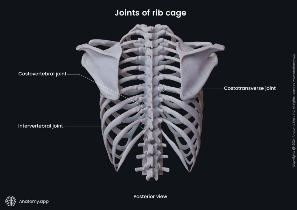
The ribs usually increase in size and length from rib 1 to rib 7, and they decrease from rib 7 to rib 12. All ribs form several articulations. Their proximal ends directly articulate with the vertebrae of the spine, forming the costovertebral joints and costotransverse joints. In contrast, the distal ends of only the first seven ribs directly articulate with the sternum.
According to their articulation with the sternum, all ribs can be grouped into three types:
- True (vertebrosternal) ribs – ribs 1 to 7; they attach directly to the sternum via their own costal cartilages;
- False (vertebrochondral) ribs – ribs 8 to 10; they are connected to the sternum indirectly via the cartilage of the rib above them; all costal cartilages of the false ribs join with the costal cartilage of the seventh rib;
- Floating (free) ribs – ribs 11 and 12; these ribs do not articulate with other ribs, adjacent costal cartilages or sternum; their costal cartilages tend to end within the abdominal musculature.
Ribs can be classified in one more way. Each rib can either be typical rib or atypical rib based on its anatomical features.
Typical ribs
The third through ninth (3 – 9) ribs share similar characteristics. They are considered typical ribs. Their main parts are the head, neck, tubercle, and body. Each part has its characteristic landmarks described below.
- Head – the most proximal portion; it appears wedge-shaped and articulates with two adjacent thoracic vertebrae;
- Articular demi-facets – two surfaces found on the head for the articulation with the corresponding thoracic vertebra and the thoracic vertebra above; the superior articular demi-facet is usually larger than the inferior one;
- Interarticular crest – a sharp wedge that separates both articular demi-facets;
- Neck – relatively short and flat segment that links the head of the rib with its body;
- Crest of the costal neck – sharp superior border of the neck that goes from the head towards the tubercle;
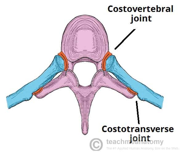
- Tubercle – a bony prominence that is found on the posteroinferior aspect of each typical rib at the junction between the neck and body; it articulates with the transverse process of the thoracic vertebra, forming the costotransverse joint; it consists of two parts:
- Non-articular part of the tubercle – rough lateral part of the tubercle that serves as the attachment site for ligaments, such as the costotransverse ligament;
- Articular part of the tubercle (articular facet) – medial part of the tubercle; it presents as a smooth surface for the articulation with the transverse process of the corresponding thoracic vertebra;
- Body (shaft) – the largest portion of the rib; it is thin, flat and curved and is found between the neck and the costal cartilage; the bodies of the third to sixth ribs are thicker and more rounded than those of the seventh to ninth ribs; it has two borders and two surfaces;
- Superior border – the upper blunt, smooth and convex edge;
- Inferior border – the sharp lower edge;
- External surface – the outer convex surface;
- Internal surface – the inner concave surface that faces the thoracic cavity;
- Costal angle – the sharp curve of the body that is formed lateral to the costal tubercle; it serves as the attachment site for some deep muscles of the back; usually, the tubercle to costal angle distance increases in the inferior direction;
- Costal groove – found along the inferior aspect of the internal surface; the ribs 5 – 7 usually have the most prominent costal grooves; it serves as the passage site for the intercostal nerve, intercostal artery and intercostal vein; these neurovascular structures are organized in a particular order – the highest is the intercostal vein, in the middle is the artery, while the nerve is located below the artery (mnemonic: VAN); please note that the intercostal nerve is usually not protected by the costal groove.
The sternal (distal) end of the rib appears porous and roughened. Sternal ends connect with the costal cartilages, forming the costochondral joints. Most of the typical ribs are directly attached to the sternum.
Note: The tenth ribs can be either typical or atypical, depending on their articulations with the bodies of the ninth to tenth thoracic vertebrae. If the tenth rib articulates with both bodies of the mentioned vertebrae, it is classified as a typical rib – its head will have two articular demi-facets. If the tenth rib articulates with only the body of the tenth thoracic vertebra (T10), it will be classified as an atypical rib. Moreover, there will be only one facet on its head.
Atypical ribs
The first rib, second rib, eleventh rib, and twelfth rib are considered atypical ribs because of their distinct characteristics reviewed in the following slides. These ribs do not have features that are usually found on all typical ribs.
Note: The tenth ribs can be either typical or atypical, depending on their articulations with the bodies of the ninth and tenth thoracic vertebrae. If the tenth rib articulates with both bodies of the mentioned vertebrae, it is classified as a typical rib – its head will have two articular demi-facets. If the tenth rib articulates with only the body of the tenth thoracic vertebra (T10), it will be classified as an atypical rib. Moreover, there will be only one facet on its head.
Atypical ribs (eleventh and twelfth ribs)
The eleventh and twelfth ribs are the most simple ribs, lacking most of the landmarks of the typical ribs. These ribs are short with narrow and pointed sternal ends. The twelfth rib is shorter than the eleventh rib and can also be shorter than the first rib.
Both ribs do not have necks and tubercles. The shaft of the eleventh rib can make a slight angle, and it can also have a shallow costal groove. In contrast, the body of the twelfth rib usually has no costal groove and angle.
The heads of the eleventh and twelfth ribs have only one articular facet, each articulating with their corresponding thoracic vertebrae.
Thoracic vertebrae
Each person has twelve thoracic vertebrae (T1 – T12). They are vertically aligned bones that form the second region of the spine, and they also participate in forming the posterior wall of the thorax. These vertebrae are positioned between the cervical vertebrae and lumbar vertebrae.
The thoracic vertebrae are characterized by their articulation with the ribs. Therefore, the main distinguishing feature of the thoracic vertebrae is the presence of the costal facets.
The thoracic vertebrae can be subdivided into typical thoracic vertebrae (T2 – T9) and atypical thoracic vertebrae (T1, T10 – T12) depending on the number and characteristics of their costal facets.
- A typical thoracic vertebra has a somewhat heart-shaped vertebral body when viewed from above and a circular vertebral foramen.
- The vertebral body of a typical vertebra has two partial facets (superior costal demi facet and inferior costal demi-facet) on each its side for articulation with the head of its corresponding rib and the head of the rib below.
- Each transverse process has a transverse costal facet that articulates with the tubercle of its corresponding rib.
The thoracic vertebrae form several joints within the thorax. Articulations between the vertebral bodies of the thoracic vertebrae and the heads of the ribs are called costovertebral joints. At the same time, the articulations between the tubercles of the ribs and the transverse processes of the thoracic vertebrae are known as costotransverse joints.
Intercostal space
The bones forming the thoracic cage are arranged in a pattern that allows some space between them. Those spaces are referred to as the intercostal spaces.
The intercostal spaces contain:
The intercostal nerves and major blood vessels lie in the costal groove along the inferior margin of the superior rib and travel between the inner two layers of intercostal muscles.
Note: In each intercostal space, the vein is the highest located structure, below which lies the artery, and the nerve lies inferiorly to the artery and often is not protected by the groove. Thus, the nerve is the structure most at risk when objects perforate the upper part of the intercostal space.
Muscles of intercostal spaces
Each intercostal space contains three layers of intercostal muscles:
- External intercostal muscle – the most superficial layer of the intercostal muscles;
- Internal intercostal muscle – lie beneath the external intercostal muscles;
- Innermost intercostal muscle – located deep to the internal intercostal muscles, mainly present in the lateral parts of the thorax.
Go to the section Thoracic wall muscles to learn more about the intercostal muscles and other muscles forming the chest wall!
Note: The 3D model is a fragment of the third intercostal space. The vein you can see going along the spine is the right superior intercostal vein.
Intercostal nerves
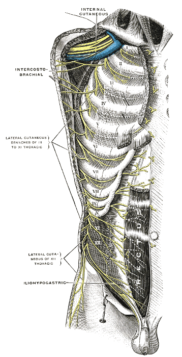
The intercostal nerves originate segmentally from the anterior rami of the thoracic spinal nerves T1 to T11. They run in the intercostal space between the adjacent ribs. The intercostal nerves are situated within or slightly below the costal grooves and go alongside the intercostal vessels. Within the subcostal groove, the intercostal nerve lies inferior to the intercostal vein and intercostal artery.
The intercostal nerves belong to the somatic nervous system and contain both – motor and sensory fibers. The efferent motor fibers supply the intercostal muscles and muscles of the anterolateral abdominal wall, while the afferent sensory fibers carry sensory information from the skin of the thoracic and abdominal walls, ribs, parietal pleura and parietal peritoneum to the brain.
At their origin sites, the intercostal nerves are connected to the sympathetic ganglion of the sympathetic trunk via the communicating branches. From the origin sites, the intercostal nerves run in the anterior direction and reach the intercostal space going between the posterior intercostal membrane and parietal pleura. The intercostal nerves enter the intercostal spaces at the angles of the ribs and further go between the innermost intercostal muscles and internal intercostal muscles.
The upper and lower intercostal nerves have different termination sites. The first six intercostal nerves (T1 – T6) end within the anterior aspects of their corresponding intercostal spaces near the sternum, while the seventh through eleventh intercostal nerves (T7 – T11) extend into the anterior abdominal wall and terminate within it. The T7 – T11 intercostal nerves are also known as the thoracoabdominal nerves.
Note: The anterior ramus of the twelfth thoracic spinal nerve (T12) is not considered an intercostal nerve. It goes below the twelfth rib towards the abdominal wall and is referred to as the subcostal nerve.
Typical and atypical intercostal nerves
All intercostal nerves can be subdivided into atypical and typical intercostal nerves depending on their course and the area they supply. The first two intercostal nerves (T1 – T2) and the lowest five intercostal nerves (T7 – T11) are classified as atypical, while the third through sixth intercostal nerves (T3 – T6) are typical intercostal nerves. Typical intercostal nerves go within their corresponding intercostal spaces, while the atypical intercostal nerves also extend into other regions and supply them.
- The first thoracic spinal nerve has two terminal branches – inferior and superior. The inferior branch forms the first intercostal nerve (T1), while the superior branch joins the brachial plexus.
- The lateral cutaneous branch of the second intercostal nerve (T2) supplies the floor of the axilla and the upper posteromedial part of the upper limb.
- The lowest five intercostal nerves (T7 – T11) terminate within the anterior abdominal wall. When reaching the costal margins, they go posterior to them, pierce the anterior rectus sheath and terminate as the anterior cutaneous branches. They supply the anterior abdominal wall and parietal peritoneum.
Branches of intercostal nerves
The intercostal nerves have several branches, and they include the following:
- Communicating branches
- Posterior branches
- Muscular branches
- Collateral branches
- Lateral cutaneous branches
- Anterior cutaneous branches
The communicating branches are found near the origins of the intercostal nerves, and they connect each intercostal nerve with the sympathetic trunk. Also, near the origin, the intercostal nerves give off posterior branches to the back muscles and their overlying skin.
The muscular branches innervate the intercostal muscles, serratus posterior superior muscle, subcostal muscles, transversus thoracis muscle and levatores costarum muscles.
The collateral branch of each intercostal nerve originates near the angle of the rib and runs along the superior border of the rib below. They innervate the intercostal muscles, parietal pleura and periosteum of the ribs.
The intercostal nerves also give off two types of cutaneous branches – lateral cutaneous branch and anterior cutaneous branch. They provide segmental sensory supply and innervate the skin of the anterolateral wall of the thorax and skin of the anterolateral wall of the abdomen.
- The lateral cutaneous branches arise near the midaxillary line of the thorax. They pierce the muscles of the lateral thoracic wall and reach the superficial soft tissues of the lateral thoracic wall, where they divide into anterior and posterior branches.
- The anterior cutaneous branches are terminal branches of the intercostal nerves. They are found at the anterior chest wall along the sternum and within the soft tissues of the anterior abdominal wall. The anterior cutaneous branches divide into the medial and lateral branches.


